
Behind Your Headaches
How the Neck-Dura Connection Triggers Pain and How to Relieve It

A muscle the size of your fingertip attaches directly to the covering of your brain. Chronic tension in this muscle produces a headache that follows the anatomy of the trigeminal nerve, so you feel it in your temple, forehead, or behind the eye. Neurologists may call it a migraine, a cervicogenic or tension-type headache.
In 1995, the space between the skull and the first vertebra of the neck was dissected and a structure that had escaped attention for more than a century was discovered. A thick band of connective tissue extended from a small muscle called the rectus capitis posterior minor (RCPmi, one of the deep suboccipital muscles at the base of the skull) directly into the spinal dura mater. The dura is the tough outer membrane covering the brain and spinal cord. Anatomy textbooks did not recognize any direct attachment between this muscle and the dura; they described the dura as floating freely inside the spinal canal. The textbooks were wrong.
The same RCPmi-dura bridge has been found in all mammals examined: a band of dense collagen fibers running from the muscle into the dura and thick enough to transfer force during muscle contraction.
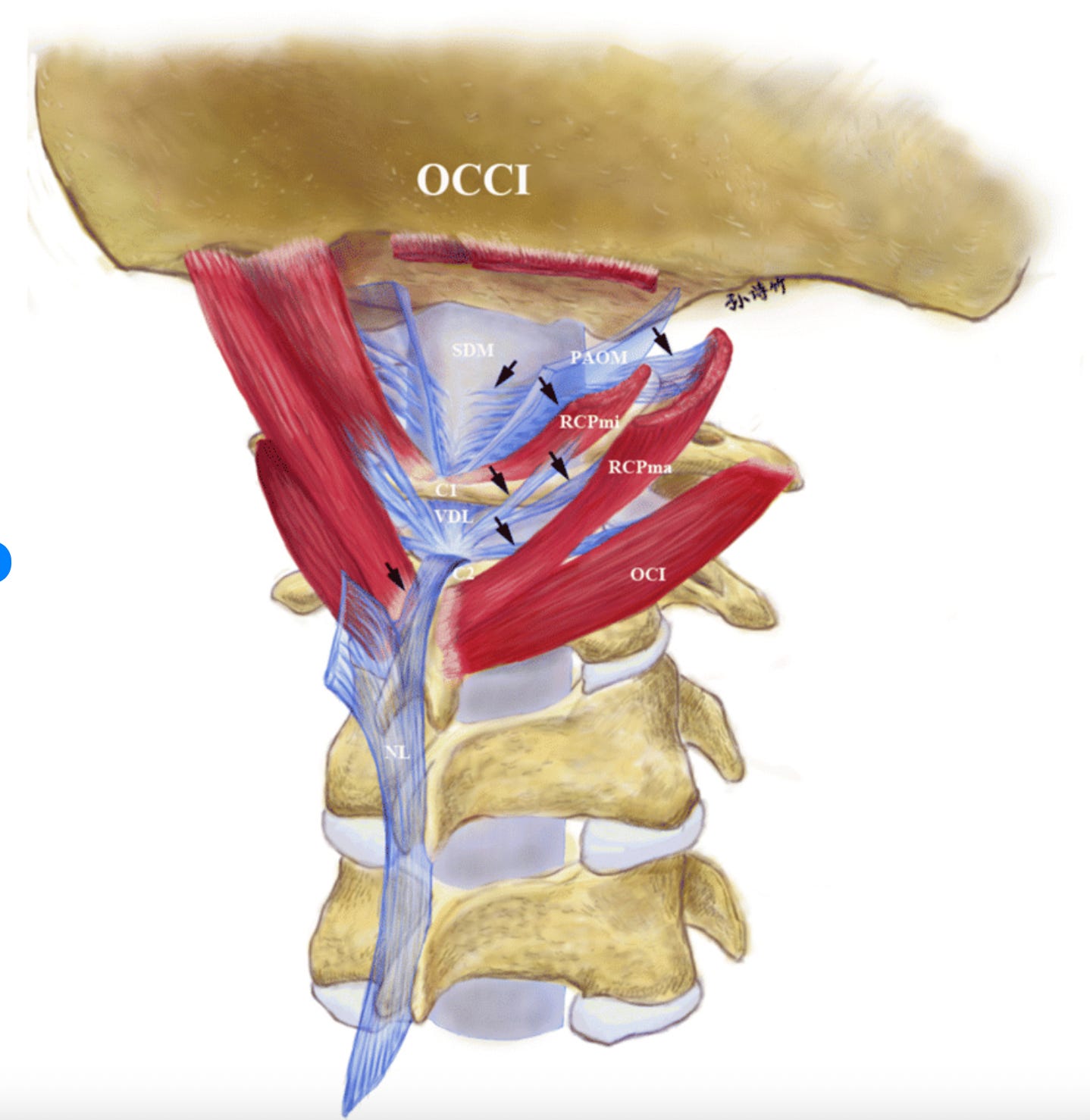
Researchers later discovered that the RCPmi wasn’t the only muscle with a connection to the dura. The rectus capitis posterior major (RCPma), a slightly larger neighboring muscle, also connects to it between the first and second cervical vertebrae. So does the obliquus capitis inferior (OCI), another muscle of the same size spanning those same two vertebrae. The nuchal ligament, a thick sheet of connective tissue running down the back of your neck, is part of the same complex too. As you can see, the dura isn’t “floating freely” inside the spinal canal. It’s anchored to the neck.
When these three muscles contract, they pull on the dura. With any head movement, that force is conveyed through the myodural bridges. Under normal circumstances, the dura moves with the muscle, and the tension is released when the muscle relaxes. Different motor units take turns firing, and blood flow increases and decreases as needed. These muscles were made for sustained, low-grade activity with plenty of variation.
This whole mechanism relies on constant head movement. If you hold your head in the same position in front of a screen for hours, the same motor units keep firing because the position doesn’t change. The tissue gets compressed, slowing the blood flow and allowing metabolic by-products to accumulate. And if the muscle stays contracted and shortened for hours, and if you have bad sleep ergonomics (pillow matters A LOT, more on that below), then you’re keeping your neck tight all night.
Because the dura is highly innervated, it responds rapidly to both mechanical strain and the resulting inflammation. On top of that, the dura is connected directly to the sensory pathways that produce head pain, and those pathways feed into the trigeminal system.
Below: How tension in the neck results in pain in the temple, forehead, or behind the eye; how mechanical input from the neck can initiate or worsen a migraine; the mechanism behind cervicogenic headaches; what craniosacral therapy gets wrong and where it may actually be helpful; and how you can help yourself at home.
The Trigeminal-Dura-Cervical Convergence
The trigeminal nerve, particularly its ophthalmic branch, V1, innervates much of the supratentorial dura and the blood vessels that surround it.
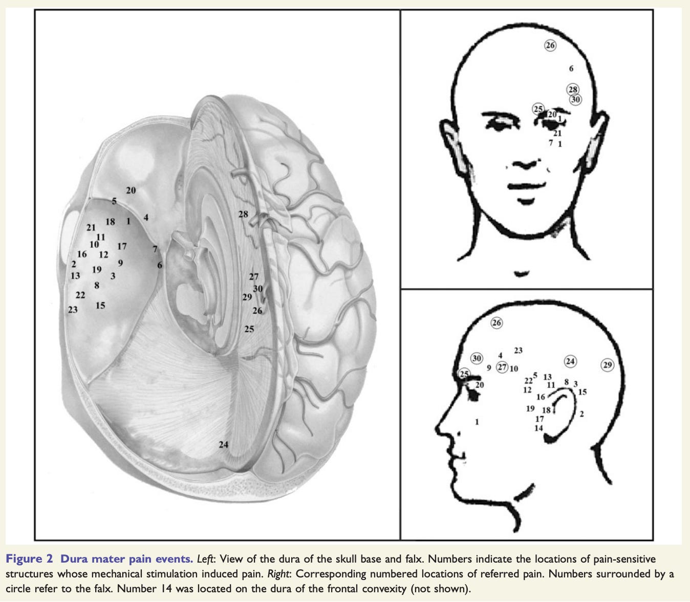
Penfield and McNaughton showed that mechanical or electrical stimulation of the dura evokes pain in the face, temple, forehead or retro-orbital region. This is called referred pain: the sensation originates at one location but is perceived in a different location that is served by the same nerve.
The trigeminal system’s sensory pathways extend down into the upper cervical spinal cord, where trigeminal input converges with sensory input from the upper cervical nerves, primarily C1–C3. Neurologists refer to this shared processing region as the trigeminocervical complex (TCC).